A sudden patch of warm, swollen skin can be deeply alarming, leaving many individuals anxious to understand whether a simple rash is rapidly escalating into a dangerous medical situation. Cellulitis is a serious, deep bacterial infection of the dermis and subcutaneous tissue—most commonly triggered by Streptococcus or Staphylococcus bacteria—that penetrates far below the outer skin layer. Because these harmful bacteria thrive beneath the surface, the infection can spread rapidly through surrounding tissue, enter the lymphatic system, and eventually reach the bloodstream if left untreated. Recognizing early visual indicators is vital to beginning target antibiotic treatment before life-threatening complications develop.
Searching online for clear pictures of cellulitis provides an invaluable visual baseline when you need to assess sudden changes in your skin tone and texture immediately. While high-quality clinical photographs help you spot key indicators like expanding borders, tight swelling, and intense localized heat, they should always serve as an educational reference rather than a definitive medical diagnosis. Using visual guides allows patients, family members, and caregivers to act quickly, recognizing when a minor skin irritation requires a prompt trip to an urgent care clinic or an immediate emergency room evaluation.
What Does Cellulitis Look Like? Early Visual Symptoms
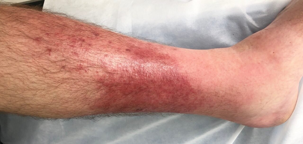
In its earliest stages, cellulitis often mimics minor skin irritation or an allergic reaction, but subtle visual cues reveal a far more aggressive process taking shape beneath the dermal layers. The affected area typically presents as a distinct, expanding section of bright redness that feels noticeably hot, tender, and firm when touched. As inflammatory fluid continuously accumulates within the deep tissue layers, the skin often stretches out, appearing tight, smooth, glossy, or swollen compared to surrounding healthy areas.
Most early infections begin at a vulnerable entry point where the natural protective skin barrier was compromised, such as a microscopic bug bite, small paper cut, surgical incision, or fungal crack between the toes. Comparing your skin to verified pictures of cellulitis can help you spot early spreading patterns before systemic symptoms emerge. A practical monitoring technique is drawing a thin pen line around the initial red border; if the redness continuously expands outside that ink mark over a few short hours, the bacterial infection is actively spreading and requires swift medical intervention.
Visual Signs Your Cellulitis Is Severe and When to Worry
When a localized bacterial infection breaches deep tissue structures, it produces distinct physical changes that signal a severe, highly dangerous medical emergency. Reviewing clinical pictures of cellulitis highlighting advanced infection stages can help you differentiate between a manageable localized infection and an aggressive outbreak demanding immediate hospital care. You must pay close attention to critical warning signs like red streaking moving toward the heart, fluid-filled blisters forming across inflamed areas, and localized yellow pus or open drainage, all of which indicate deep tissue involvement requiring systemic intravenous antibiotic treatment.
Furthermore, severe infections often cause the affected skin to transform from a bright red shade into a dark purple, gray, or mottled black color, signaling localized tissue death known as necrosis. This rapid discoloration is a hallmark sign of necrotizing fasciitis, an extremely dangerous condition that demands emergency surgical intervention to remove dead tissue. When rapidly expanding red margins are accompanied by systemic visual distress—such as high fevers, severe chills, cold sweats, or mental confusion—it indicates that the bacterial infection has likely entered the bloodstream, evolving into life-threatening sepsis.
Cellulitis by Body Location: Legs, Feet, Face, and Arms

Cellulitis can develop virtually anywhere on the human body, but it most frequently strikes the lower legs and feet due to constant gravitational pressure, reduced circulation, and undetected minor injuries. Lower extremity infections often cause dramatic swelling around the ankles and shins, making walking extremely painful as fluid pools within the soft tissues. Examining real pictures of cellulitis on lower limbs demonstrates how chronic underlying conditions like venous insufficiency, lymphedema, or athlete’s foot create perfect microscopic entryways for environmental bacteria to invade deep tissue layers.
Infections affecting the face, hands, or arms carry heightened physiological risks that demand immediate clinical evaluation from an experienced healthcare professional. Facial cellulitis—particularly around the eyes, cheeks, or nose—can quickly travel to deeper cranial structures, potentially impacting vision or triggering serious central nervous system infections. On the arms and hands, tight anatomical spaces mean localized tissue swelling can compress delicate nerves and blood vessels, frequently occurring after animal bites, deep puncture wounds, or intravenous catheter insertions.
Visual Distinctions: Is It Cellulitis or Something Else?
Diagnosing skin conditions purely by visual appearance can be challenging because several distinct medical conditions closely mimic the localized redness, warmth, and swelling of bacterial skin infections. For instance, erysipelas is a closely related superficial skin infection that displays bright red, raised lesions with sharp, distinctly elevated borders, whereas cellulitis features deeper, flatter redness with blurred, poorly defined margins. Looking closely at direct side-by-side pictures of cellulitis alongside other skin conditions reveals these distinct structural differences in skin texture, elevation, and border clarity.
Other non-infectious medical issues are frequently mistaken for active bacterial infections, leading to improper treatment plans and unnecessary antibiotic use. Stasis dermatitis causes bilateral redness and scaly skin on both lower legs due to failing vein valves, unlike cellulitis which almost always appears on just one leg. Similarly, Deep Vein Thrombosis (DVT) creates deep leg pain, intense warmth, and heavy localized swelling caused by a blood clot inside a deep vein, but it lacks the bright, spreading surface rash characteristic of true bacterial cellulitis.
Causes, Risk Factors, and Common Triggers
Cellulitis develops when environmental bacteria successfully break through the protective outer epidermis and multiply within the deeper dermal and subcutaneous tissues. Group A Streptococcus and Staphylococcus aureus are the primary bacterial culprits responsible for these aggressive soft-tissue infections. Even seemingly trivial entry points like dry, cracked heels, minor insect stings, scratch marks, or persistent athlete’s foot fungal infections provide ideal doorways for bacteria to establish themselves deep beneath the skin surface.
Certain underlying health factors significantly increase your personal susceptibility to developing severe or recurring bouts of cellulitis over time. Individuals living with diabetes, chronic kidney disease, active cancer treatments, or compromised immune systems face substantially higher infection risks and much slower healing rates. Furthermore, chronic conditions that impede normal fluid drainage and blood circulation—such as lymphedema, venous insufficiency, varicose veins, or severe leg swelling—create prime biological conditions for bacteria to rapidly multiply beneath the skin.
What to Do Next: Medical Diagnosis, Treatment, and Prevention
If you notice spreading skin redness accompanied by heat, tightness, and localized pain, seeking a medical evaluation is essential to prevent deeper tissue complications. Mild, highly localized infections without systemic fever can typically be diagnosed at an urgent care clinic, where doctors will prescribe a course of targeted oral antibiotics. However, if you develop high fevers, red streaking, purple skin discoloration, or intense pain out of proportion to the injury, seek emergency room care immediately, as IV antibiotics and hospital monitoring may be required.
Preventing future recurring bacterial skin infections relies on maintaining an intact skin barrier and practicing diligent wound care every single day. Wash any minor cuts, bug bites, scrapes, or punctures immediately with mild soap and clean water, applying a sterile protective bandage until the surface fully heals. Additionally, keeping your skin thoroughly moisturized to prevent painful cracking, treating fungal foot infections promptly, and managing chronic leg swelling with prescribed compression garments under medical supervision will significantly lower your overall infection risk.
Conclusion
While reviewing authentic pictures of cellulitis offers essential visual guidance for spotting early skin changes, recognizing severe warning signs like red streaking, dark purple skin discoloration, and blistering is what truly saves lives. Because deep bacterial infections escalate rapidly, relying solely on self-evaluation online can lead to dangerous delays in receiving professional care. Always consult a qualified healthcare provider at the first sign of spreading skin redness, localized heat, or systemic fever to ensure you receive proper antibiotic treatment and protect your overall health.
Frequently Asked Questions (FAQs)
What does cellulitis look like when it first starts?
Early cellulitis typically appears as a small, flat patch of red, tender, and slightly swollen skin. It feels noticeably warmer than the surrounding tissue and usually spreads outward over time from a visible cut, insect bite, or microscopic break in the skin barrier.
How quickly does cellulitis spread across the skin?
Cellulitis can spread within a few hours or over a couple of days, depending on the specific bacteria involved and the strength of the individual’s immune system. Rapidly expanding redness accompanied by fever, chills, or red streaking is a serious medical emergency.
Can pictures of cellulitis help me self-diagnose at home?
Pictures of cellulitis provide a helpful visual reference to compare your symptoms, but they cannot replace a direct medical evaluation. Other medical issues like DVT, stasis dermatitis, and severe allergic reactions look very similar and require completely different treatments.
Is cellulitis contagious through direct contact or looking at it?
Cellulitis itself is not contagious through casual contact because the infection resides deep within the lower skin layers. However, if there is open pus or active fluid drainage, the bacteria inside that fluid can spread to others through direct contact with unwashed hands or broken skin.
What is the main difference between mild cellulitis and severe cellulitis?
A: Mild cellulitis presents with localized skin redness, slight swelling, and light tenderness without any systemic fever. Severe cellulitis involves dangerous systemic symptoms like high fevers, rapid red streaking, fluid-filled blisters, or skin turning dark purple or black.
You may also read : Real Health Benefits You Can Expect