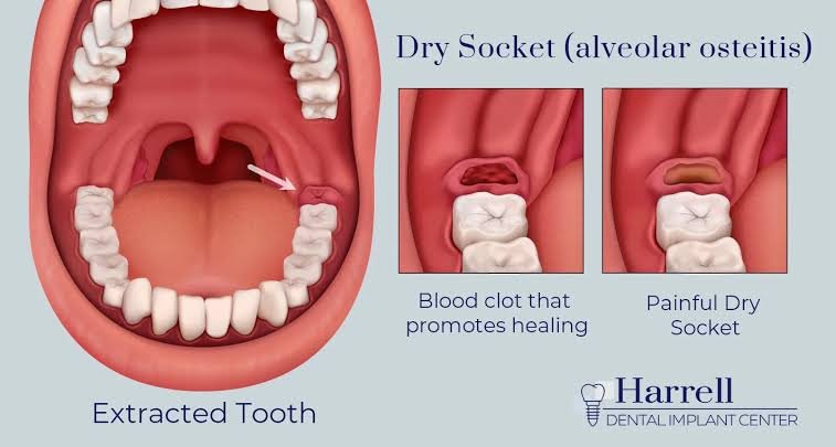
Recovering from a tooth extraction can feel like a stressful waiting game filled with uncertainty and frequent visual checks. Immediately after a dentist or oral surgeon removes a tooth—whether a simple molar or an impacted wisdom tooth—your body naturally initiates a complex tissue repair process. The single most crucial event in this initial recovery stage is the formation of a dark red blood clot inside the open extraction cavity. This vital clot protects delicate nerve endings and underlying jawbone while creating a biological foundation for new soft tissue to grow smoothly over the coming weeks.
Because the back of the mouth is dark, tight, and difficult to inspect without good lighting, many patients regularly consult pictures of dry socket vs normal healing to make sense of what they see in the mirror. Reviewing accurate reference images helps you systematically monitor expected recovery milestones while spotting early warning signs of alveolar osteitis, commonly referred to as a dry socket. Understanding what healthy healing looks like brings peace of mind, whereas recognizing how a dry socket behaves ensures you seek timely professional intervention before discomfort turns into unbearable pain.
Visual Comparison: Normal Healing vs. Dry Socket (Day-by-Day Timeline)
On the first day following surgery, a healthy socket features a deep crimson, jelly-like blood clot that completely fills the void left by the extracted tooth. This primary seal shields sensitive nerves from air, fluids, and food particles. If you engage in risky behaviors such as smoking, aggressive mouth rinsing, or drinking through a straw during these critical first twenty-four hours, you dramatically increase your risk of dislodging or dissolving this fragile blood clot before it secures itself to the surrounding bone walls.
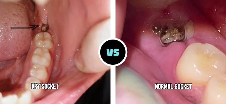
By the second and third days, a normal socket begins developing a whitish or cream-colored layer of granulation tissue directly over the dark clot. Patients who inspect their mouths often panic, mistaking this healthy, collagen-rich repair tissue for pus or a bacterial infection. However, comparing pictures of dry socket vs normal healing during this stage reveals that a dry socket lacks this protective layer entirely. Instead, it presents as a dark, empty hole or reveals a dull, off-white surface of exposed jawbone that remains completely unprotected.
During days four through seven, normal recovery causes the physical gap to shrink significantly as soft tissue steadily coats the inner wound walls and post-operative soreness fades away. Conversely, a dry socket reaches its peak severity during this mid-recovery window. Instead of experiencing steady relief, patients suffer intense, throbbing pain that frequently radiates toward the ear, temple, or neck, accompanied by a static, hollow-looking socket that shows zero signs of progressive tissue coverage or visual improvement.
From day eight through fourteen, a healthy extraction site exhibits smooth, light-pink mucosal tissue that gradually closes the opening, leaving only a shallow indentation in the gumline. If you developed a dry socket and received proper clinical care with medicated dressings, your visual recovery timeline will simply lag behind by several days. The socket eventually fills with healthy tissue once local inflammation subsides, allowing your gums to restore their natural structure without long-term structural complications.
Key Visual and Physical Differences (Side-by-Side Breakdown)
Color variations offer one of the most reliable visual markers when assessing the condition of your extraction site day by day. A healthy healing socket transitions naturally from a dark red clot to a pale, yellowish-white shade as new blood vessels and connective tissues form. In contrast, examining pictures of dry socket vs normal healing demonstrates that a dry socket fails to show this healthy color transition, displaying either a dark, empty crater or a dry, grayish-white bone bed that completely lacks healthy vascular coverage.
Structural appearance further clarifies the distinction between a normal recovery path and alveolar osteitis. A healthy socket appears filled, moist, and slightly recessed, with the surrounding gum tissue pulling inward to seal the opening. A dry socket looks noticeably hollow and vacant, resembling a deep pocket where the essential blood clot vanished. Beyond visual evidence, physical symptoms confirm the diagnosis: normal healing produces mild, manageable soreness, whereas a dry socket causes severe throbbing pain, foul breath, and a persistent metallic or bitter taste.
Causes and Risk Factors for Dry Socket
A dry socket develops when the blood clot either fails to form properly, gets mechanically dislodged, or dissolves prematurely due to chemical breakdown before tissue repair occurs. Daily habits during early recovery frequently trigger this complication. Using drinking straws creates negative suction inside the oral cavity, while forceful spitting, vigorous saltwater rinses, or repeatedly probing the socket with your tongue can physically dislodge the delicate clot from the underlying bone wall.
Chemical contaminants and underlying health factors significantly heighten the probability of developing alveolar osteitis post-surgery. Inhaling tobacco smoke or vaping aerosol introduces harmful chemicals that constrict micro-blood vessels and stall natural tissue repair, while the physical inhalation force adds dangerous suction. Furthermore, existing gum disease, elevated estrogen levels from oral contraceptives, and complex surgical extractions—particularly lower impacted wisdom teeth—statistically increase the overall incidence of dry socket complications in adult patients.
Prevention, Home Care, and Medical Treatment
Preventing a dry socket requires strict adherence to your dental surgeon’s post-operative recovery protocol for at least seven days. Avoid using drinking straws, smoking cigarettes, or vaping for three to seven days post-surgery to maintain stable pressure within your mouth. Keep adjacent teeth clean through gentle brushing, but avoid rinsing aggressively. Stick exclusively to soft, lukewarm foods such as yogurt, smooth soups, and mashed vegetables, always eating with a spoon to protect the surgical site.
To support healthy tissue regeneration during normal recovery, gently bathe the area with warm saltwater starting twenty-four hours after your procedure. Manage typical post-operative discomfort with prescribed or over-the-counter anti-inflammatory medications like ibuprofen. If your symptoms match pictures of dry socket vs normal healing, contact your dental office immediately. A dentist can gently irrigate food debris from the cavity and apply a specialized medicated paste containing eugenol to provide rapid, targeted pain relief.
Conclusion
Distinguishing between expected surgical recovery and painful post-operative complications becomes straightforward when you consult pictures of dry socket vs normal healing. A healthy healing socket showcases a dark red clot that gradually evolves into whitish granulation tissue while discomfort steadily decreases. On the other hand, a dry socket appears as an empty, bone-revealing cavity characterized by intense, radiating pain that worsens several days after an otherwise uncomplicated extraction procedure.
While visual self-inspections offer valuable context, your subjective pain level remains the most accurate indicator of your underlying recovery status. Normal healing discomfort responds well to mild pain relievers and diminishes steadily each day. If your pain intensifies, spreads across your jaw or ear, or fails to respond to standard medication, do not rely solely on online photographs—contact your dental clinician promptly for a professional evaluation and effective pain management.
Frequently Asked Questions (FAQs)
What does a normal healing socket look like on Day 3?
By the third day after an extraction, a normal healing socket contains a stabilized dark blood clot partially covered by pale, cream-colored granulation tissue. The surrounding gums may still display mild redness or swelling, but the socket itself should look moist, filled, and protected rather than hollow or empty.
What is the white stuff inside my tooth socket?
In the majority of cases, white or yellowish tissue inside a healing socket represents normal granulation tissue composed of collagen, new capillaries, and white blood cells repairing the area. However, if the white surface appears hard, dry, and accompanied by severe throbbing pain, it likely indicates exposed jawbone from a dry socket.
Can you have a dry socket without severe pain?
While uncommon, mild cases involving partial loss of the blood clot can produce minor discomfort rather than agonizing pain. However, classic alveolar osteitis is almost universally characterized by intense, radiating pain that spreads to the ear, eye, or temple between two and four days after the surgical extraction.
How do I know if the blood clot came out of my socket?
You will typically notice an empty gap or visible dark hole where the tooth was removed, occasionally seeing dark tissue fragments when spitting. Within twenty-four to forty-eight hours of losing the protective clot, you will experience a dramatic surge in localized and radiating pain throughout the affected jaw region.
How long does the risk of dry socket last after tooth extraction?
The highest risk window for developing a dry socket occurs during the first three to four days following your procedure. By days seven to ten, newly formed granulation tissue securely coats the exposed bone, and the risk of developing alveolar osteitis drops to practically zero.
You may also read: Step-by-Step Guide on How to Book Specsavers Ear Wax Removal Online